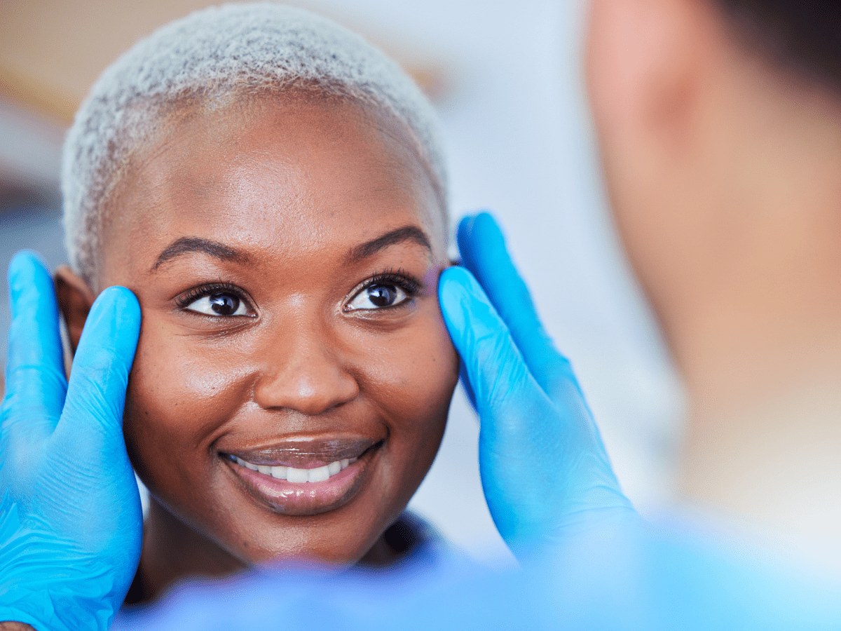
What Is Skin Cancer?
Skin cancer is a type of cancer that develops in the cells of the skin. It occurs when skin cells grow abnormally and uncontrollably, often due to DNA damage caused by factors like ultraviolet (UV) radiation from the sun or tanning beds.
Basal Cell Carcinoma
This type of cancer is the most common and the most treatable variety. Experts estimate that about 2.8 million cases of basal cell carcinoma are discovered every year in the U.S. Two factors contribute to the development of this type of cancer: a genetic predisposition and excessive sun exposure. It’s typical for people to have this illness without realizing it, which is why total body skin exams are so important.
Squamous Cell Carcinoma
Squamous cell cancer is the second most prevalent type of skin cancer. In the United States, about 700,000 people are diagnosed with squamous cell every year. Squamous cell is also curable in many cases. Like basal cell, sun exposure and a genetic predisposition are common factors for this type of cancer. Sometimes squamous cell makes itself known with a lesion that itches or bleeds. Left untreated, the lesion can metastasize under the skin to spread. Patients should watch lesions carefully. Anytime a sore doesn’t heal, see a physician to have it examined.
Malignant Melanoma
Malignant melanoma is the most deadly type of skin cancer. Although less common, melanoma claims over 9,000 lives per year in the U.S. Every year, over 76,000 new cases of melanoma will be diagnosed in the U.S. UV light exposure is a common cause of melanoma. Atypical moles can increase someone’s risk of developing melanoma. Anyone with a high number of irregular or large moles should have a total body skin exam regularly.
Others
Kaposi’s sarcoma is a rare cancer that is a result of Human Herpes Virus 8. This type of cancer results in lesions on the legs and feet in brownish-red or blue colors. Actinic Keratosis is a condition that occurs prior to squamous cell carcinoma. With this issue, lesions will develop that are scaly, rough, and red or pink in color.
Understanding types of skin cancer will allow you to monitor your own health proactively. Contact us today for more information!

FAQs
How do I recognize Basal Cell Carcinoma?
Basal Cell Cancer can present in a multitude of ways. Often a nonhealing lesion can be the earliest sign of a newly formed tumor. Bleeding, pain, tingling, and itching can also be very early symptoms. Tumors can either appear as an inconspicuous red bump with a central dimple or depression or a simple benign flat red region on the skin. When an ulcer or crust is present, bleeding is often a common complaint.
If a non-healing lesion such as those described above occurs on your skin, it is advisable to have your dermatologist assess the area as soon as possible, especially if you have had extensive sun exposure or a personal or family history.
Does Basal Cell Carcinoma spread internally?
This question is often asked and can be divided into two parts. First, the good news. This type of cancer does not usually metastasize or spread to other organs. The second aspect of this question, however, recognizes that these tumors can become a destructive force. Basal Cell cancer, if left untreated, can become invasive and grow deep into the skin and underneath structures.
This process can result in a disfiguring appearance and even the destruction of a sensory organ such as the ear, eye or nose. Therefore, especially on the on the head and neck areas, these cancers need to be treated early and aggressively.
What are the warning signs of skin cancer?
Signs of skin cancer include:
- A new growth or sore that doesn’t heal.
- Changes in an existing mole (size, color, shape).
- Itching, tenderness, or pain in a mole or skin lesion.
- Rough, scaly patches of skin.
- A shiny bump, red spot, or scar-like area on the skin.
What does melanoma look like?
Melanoma often looks like an irregularly shaped or multicolored mole. The “ABCDE” rule helps identify suspicious moles:
- A: Asymmetry (one half doesn’t match the other).
- B: Border (irregular, scalloped, or poorly defined edges).
- C: Color (varied colors like brown, black, red, white, or blue).
- D: Diameter (larger than a pencil eraser, although smaller melanomas can be dangerous).
- E: Evolving (changing in size, shape, or color over time).
Our Locations
Visit Dermatology Institute
Our team provides thoughtful, expert care for all your skin health needs. We are proud to offer the most advanced general, surgical, and cosmetic dermatological services in the Newnan and LaGrange areas.
Sun Protection Tips
The following is a list of commonly suggested recommendations by dermatologists to help minimize excessive sun exposure and the risk of associated skin cancer development. The American Academy of Dermatology and American Cancer Society have come up with certain recommendations to follow. Remember that every sunburn that you receive induces a costly skin damaging event. Over several decades, these effects become cumulative – resulting in skin cancer formation, wrinkles, pigment abnormalities, easy bruising and… yes, oily skin. You can “Fry now and Pay later” or maybe try to follow a more skin conscious lifestyle. Let’s try to get you started on a safe sun program by following the rules listed below.
1.Minimize sun exposure between the hours of 10 AM and 4 PM.
This is the time when the sun is at its highest level in the sky. Take shade, and think about rescheduling that jog or tennis match either early in the day or after work, especially in the summer months.
2.Don’t use Tanning Beds.
These can give you 15 times the exposure to dangerous UVA and UVB rays, which can promote melanoma development. Tanning Bed use is one reason that we are seeing an increase in melanoma development in younger women. In addition, tanning beds enhance the the rate that your skin ages, specifically causing wrinkles to develop earlier. Consider the use of self tanners instead. They are safer.
3.Use a sunscreen with at least an Sun Protection Factor of 30 or higher that says Broad Spectrum.
A 30 SPF sunscreen gives you about 96 to 97 % protection from the sun. If you are involved in an outdoor activity where you are sweating or in the pool-use a clear gel base sunscreen for this will not sweat off as easy. Sunscreens of this type should say “water resistant”. The FDA is now mandating that all sunscreens say how long they protect, e.g., sweat resistant 40 or 80 minutes. Lotion and cream based sunscreens are fine for everyday use but don’t rely on them for recreational purposes. Remember to apply the sunscreen 15 to 20 minutes before you venture outdoors. Look for sunscreens that contain Helioplex™, Mexoryl™ or Avobenzone.
Skin Cancer Facts
- Skin cancer is the most common form of cancer in the United States. More than 3.5 million skin cancers in over two million people are diagnosed annually.1
- Each year there are more new cases of skin cancer than the combined incidence of cancers of the breast, prostate, lung and colon.2
- Treatment of nonmelanoma skin cancers increased by nearly 77 percent between 1992 and 2006.
- Over the past three decades, more people have had skin cancer than all other cancers combined.3
- One of the more troubling skin cancer facts is that one in five Americans will develop skin cancer in the course of a lifetime.5
- 13 million white non-Hispanics living in the US at the beginning of 2007 had at least one nonmelanoma skin cancer, typically diagnosed as basal cell carcinoma (BCC) or squamous cell carcinoma (SCC).3
- Basal cell carcinoma is the most common form of skin cancer; an estimated 2.8 million are diagnosed annually in the US. BCCs are rarely fatal, but can be highly disfiguring if allowed to grow.6
- Squamous cell carcinoma is the second most common form of skin cancer. An estimated 700,000 cases of SCC are diagnosed each year in the US.6,7
- An estimated 3,170 deaths from nonmelanoma skin cancers will occur in the US in 2013.2
- Between 40 and 50 percent of Americans who live to age 65 will have either BCC or SCC at least once.
- Actinic keratosis is the most common precancer; it affects more than 58 million Americans.8
- Approximately 65 percent of all squamous cell carcinomas and 36 percent of all basal cell carcinomas arise in lesions that previously were diagnosed as actinic keratoses.9
- About 90 percent of nonmelanoma skin cancers are associated with exposure to ultraviolet (UV) radiation from the sun.10
- Half of all adults report at least one sunburn in the past 12 months